How Does the Heart Work?
Reviewed by: HU Medical Review Board | Last reviewed: October 2019 | Last updated: March 2021
The heart is a muscular organ about the size of an adult fist.1 It sits just to the left of the center of the chest, also known as the thoracic cavity. It beats around 100,000 times in a day and moves a total of 5,000 gallons of blood throughout the body each day.1,3 Each person has about 8 pints of blood in their body that travels through the heart.
The heart is the focal point of the circulatory system. It pumps oxygen, water, and nutrient-rich blood out to the tissues and removes waste from the bloodstream throughout the body.4 Its two main pathways are called arteries and veins. Arteries carry oxygenated blood away from the heart. Veins carry blood back to the heart where it can be sent to the lungs to be oxygenated again.5
Structure of the heart
The heart has four chambers, two upper and two lower. The upper chambers are called atria and the lower chambers are called ventricles. The chambers are labeled for the side of the body they are on, the left or the right (left atrium and right atrium, left ventricle and right ventricle). When the heartbeats, it contracts and relaxes, first squeezing the blood out of the chamber and then relaxing to refill with blood. As the heart pumps blood, it goes through a sequence of contractions. For the heart to work properly, it relies on each of the chambers to beat in an organized way.4
Figure 1. Diagram of basic heart structure
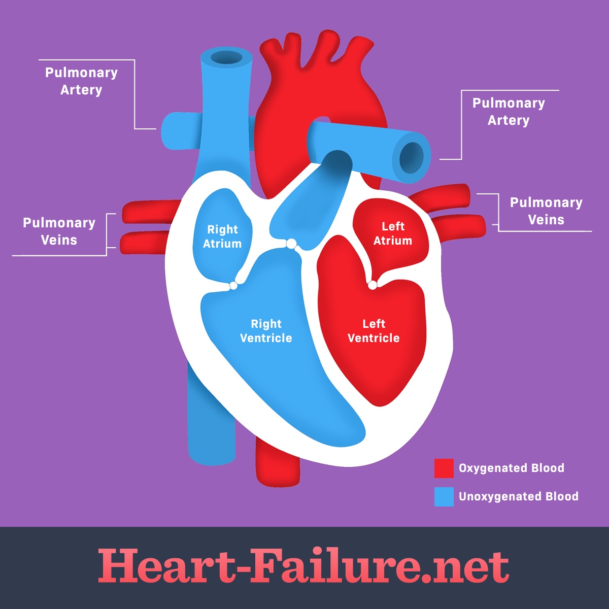
Oxygen-rich blood, which is bright red, enters the heart in the left atrium through the pulmonary vein. It then passes into the left ventricle, which pumps it out to the rest of the body. After it has traveled through the body it returns to the heart through the right atrium. From there it sends the oxygen-depleted blood back into the lungs through the right ventricle where the blood again becomes oxygenated.1
Coronary arteries
Coronary arteries supply blood to the heart muscle. They are wrapped around the outside of the heart.6 Problems with the arteries can reduce the flow of oxygen and nutrients to the heart muscle which can lead to coronary artery disease, heart attack, or even death. The aorta is the largest artery in the body and the main transport system that branches off into smaller arteries to supply the whole body with oxygen-rich blood.5
The 2 main coronary arteries are the left main and right coronary arteries. The left main coronary artery (LMCA) supplies blood to the left ventricle and left atrium of the heart. The left main coronary divides into two branches. The left anterior descending artery branches off the left coronary artery and supplies blood to the front of the left side of the heart while the circumflex artery branches off and wraps around the heart. The circumflex artery supplies blood to the outer side and back of the heart. This side of the heart is larger and more muscular because it pumps blood throughout the body.7
The right coronary artery (RCA) is smaller than the left side. It supplies blood to the right ventricle, the right atrium, and 2 nodes that regulate the heart rhythm. The nodes are called the SA (sinoatrial) node and AV (atrioventricular) node. Their job is to generate an electrical signal which tells the heart to contract. The SA node is the pacemaker and signals the upper chambers, the atria; and the AV node transmits the signal to the lower chambers, the ventricles. The right coronary artery also divides into smaller arteries, including the right posterior descending artery and the acute marginal artery. The middle of the heart, known as the septum, is supplied by both the right coronary artery and the left anterior descending artery.6,7
Composition of the heart
The heart wall is made up of three layers of tissue which are encased in a thin protective sac covering called the pericardium.8 The pericardial sac contains fluid which provides lubrication to the surface of the heart permitting freedom of movement while the heart pumps. The three layers are:
- Epicardium (a protective layer of mostly connective tissue)
- Myocardium (the muscles of the heart)
- Endocardium (the lining of the heart which protects the valves and chambers)
The heart also has four valves. The mitral valve and tricuspid valves regulate blood flow from the left and right atria respectively into the ventricles. The aortic valve and pulmonary valve control the flow of blood out of the left and right ventricles into the aorta and pulmonary artery. Heart valves direct the smooth flow of blood in one direction. When fully functioning, they quickly open and close, preventing the blood from flowing back through the valve.9
Pathophysiology of heart failure
Pathophysiology refers to the functional changes that accompany a specific disease. Heart failure means that the heart muscle isn’t pumping as well as it should be. This is called cardiomyopathy. There are functional changes in the physical structure of the heart that are associated with aging and the reduction of cardiac output. The effects result in the body tissue not getting an adequate blood supply to carry out its metabolic function. This can result in you being tired, short of breath, and coughing. Everyday activities like walking, climbing stairs, or carrying heavy packages can become more difficult.1